NIST Develops New Nondestructive Method for Assessing Bioengineered Artificial Tissues
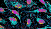
 Engineering organs to replace damaged hearts or kidneys in the human body may seem like something out of a sci-fi movie, but the building blocks for this technology are already in place. In the burgeoning field of tissue engineering, live cells grow in artificial scaffolds to form biological tissue. But to evaluate how successfully the cells develop into tissue, researchers need a reliable method to monitor the cells as they move and multiply.
Now, scientists at the National Institute of Standards and Technology (NIST), the U.S Food and Drug Administration (FDA) and the National Institutes of Health (NIH) have developed a noninvasive method to count the live cells in a three-dimensional (3D) scaffold. The real-time technique images millimeter-scale regions to assess the viability of the cells and how the cells are distributed within the scaffold — an important capability for researchers who manufacture complex biological tissues from simple materials such as living cells.
|