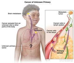
Carcinoma of unknown primary; drawing shows a primary tumor that has spread from an unknown site to other parts of the body (the lung and the brain). An inset shows cancer cells spreading from the primary cancer, through the blood and lymph systems, to another part of the body where a metastatic tumor has formed.
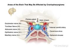
Drawing showing a coronal view of the inside of the brain where craniopharyngiomas may form. A tumor is shown between the pituitary gland and the optic chiasm. Also shown is the oculomotor nerve (III), trochlear nerve (IV), abducens nerve (VI), ophthalmic nerve (V1), maxillary nerve (V2), internal carotid artery, and the cavernous and sphenoid sinuses.
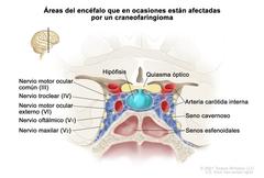
En la imagen se observa una vista frontal del interior del encéfalo donde se forman los craneofaringiomas. Se observa un tumor entre la hipófisis y el quiasma óptico. Además, se muestra el nervio motor ocular común (III), el nervio troclear (IV), el nervio motor ocular externo (VI), el nervio oftálmico (V1), el nervio maxilar (V2), la arteria carótida interna, un seno cavernoso y los senos esfenoidales.