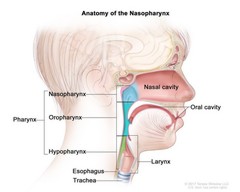
Anatomy of the nasopharynx; drawing shows the three parts of the pharynx (throat): the nasopharynx, oropharynx, and hypopharynx. Also shown are the nasal cavity, oral cavity, larynx, esophagus, and trachea.
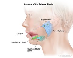
Anatomy of the salivary glands; drawing shows a cross section of the head and the three main pairs of salivary glands. The parotid glands are in front of and just below each ear; the sublingual glands are under the tongue in the floor of the mouth; and the submandibular glands are below each side of the jawbone. The tongue and lymph nodes are also shown.
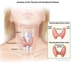
Anatomy of the thyroid and parathyroid glands; drawing shows the thyroid gland at the base of the throat near the trachea. An inset shows the front and back views. The front view shows that the thyroid is shaped like a butterfly, with the right lobe and left lobe connected by a thin piece of tissue called the isthmus. The back view shows the four pea-sized parathyroid glands. The larynx is also shown.
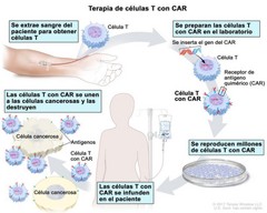
Terapia de células T con CAR; en el dibujo se muestra que se extrae sangre de una vena del brazo de un paciente para obtener células T. También se muestra que se prepara en el laboratorio un receptor especial, que se llama receptor de antígeno quimérico (CAR); se inserta el gen que produce el CAR en las células T y luego se reproducen millones de células T con CAR. Además, se muestra que el paciente recibe las células T con CAR por infusión, y que luego estas células se unen a los antígenos de las células cancerosas y las destruyen.