Potential harmful algal bloom exposure this summer in pets and livestock
Dr. Diane Gerken, DVM, ADDL Toxicologist
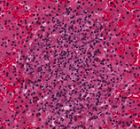
Blue-green algae blooms in ponds, lakes and standing
water are predicted to occur in Ohio again this year which means the potential
for serious and sometimes deadly health effects in large/small animals and
humans. Blue-green algae have the potential to produce many toxins – the
two most commonly found are microcystins and anatoxin A. Ingestion of
microcystins results in severe liver disease (clinical signs include lethargy,
vomiting, diarrhea, weakness and pale mucous membranes) and often death.
Anatoxin A ingestion results in central nervous system effects (clinical
signs include muscle tremors, rigidity, lethargy respiratory distress and
convulsions) often resulting in death. Gastrointestinal contents (or rumen
contents) can be analyzed for either of these toxins using submitted samples or
whole animal submission for necropsy to the ADDL. Whether it is a pet or
farm animal that is affected, a positive diagnosis is recommended so
steps to prevent additional animal or human exposure can be instituted as soon
as possible.
In addition, detection of microcystins in water
is possible using an ELISA-based assay. For testing of water sources, a list of
laboratories can be found on the EPA website. Find information about pets and harmful algal bloom exposure here.
 Comparison of water without (left) and with (right) blue green algae present.
Dr. Jeff Hayes, DVM, MS, ADDL Pathologist
Francisella tularensis, the causative agent
of the disease tularemia, was isolated from the liver of an adult female gray
fox that was found dead in Gallia County in February, 2016. External
examination showed generalized muscle wasting, marked dehydration, and a
moderate number of fleas in the hair coat. Necropsy showed scant adipose
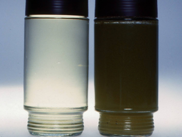
tissue, gastroenteritis including gastric ulcers, and mild enlargement of mesenteric lymph nodes. Histopathology revealed <1 mm
multifocal necrotizing lesions in the liver (too small to visualize grossly),
and widespread necrosis in the cortex of the mesenteric lymph node. Microscopic
changes were consistent with tularemia, so culture of liver tissue was
performed. F. tularensis was isolated from the liver at the ADDL,
corroborated by positive MALDI-TOF identification and PCR results, and
was confirmed as F. tularensis by the National Veterinary Services
Laboratories (NVSL).
Francisella tularensis can infect a wide
range of animals (>190 mammalian, 23 avian, 3 amphibian and 88 invertebrate
species) , including a report of infection in wild gray foxes in Minnesota
(Schlotthauer et al, Journal of Infectious Diseases, 1935). However, it
primarily causes disease in rabbits and rodents. This is the first isolation of
this organism at the ADDL since 2010, when isolations were made from two cats
and a cottontail rabbit in Montgomery County, Ohio. Transmission of tularemia
can occur by various routes, such as by the bites of arthropods and insects, including
mosquitoes, fleas, ticks, deer flies; by contact with blood or tissues of
infected animals through the skin (intact or lacerated); through
conjunctival membranes; by inhalation; or by ingestion of contaminated meat and
water.
The fox was found to have
several other infections, including canine distemper virus, intestinal Taenia
and Dipylidium tapeworms as well as nematodes, and Demodex mites
within hair follicles. Finding several typical endemic disease and parasite
agents does not rule out the need for thorough laboratory examination to detect
disease agents that can cause serious disease in humans. This case points out
the need to take care when handling any sick or dead animal, particularly
wildlife.
Focus of necrotizing hepatitis in an adult female gray fox (200X magnification)
 Top row, L to R: Dr. Alice Roudabush, Scott Fox, Dr. Jeff Hayes, Shawn Smith, Dr. David Newman; Bottom row, L to R: Amanda Gillard, Ali King, Dr. Craig Sarver
Ali King, ADDL Veterinary Pathology Assistant
The Pathology department is comprised of four
Veterinary Pathologists, two Veterinary Pathology Assistants, and two
Histotechnicians. Our pathologists specialize in the diagnosis of
diseases, through necropsy examinations of fresh and fixed animal tissues.
Our department addresses many components of disease in multiple animal species
and birds which include causes of illness and death, biopsies, morphologic
changes, in animals both living and dead. Our histotechnicians
prepare the microscopic tissues slides, and perform specialized tissues stains
and immunohistochemistry to aid in diagnostics.
Increased incidence of swine influenza (SIV)
Melanie Prarat Koscielny, ADDL Virology Laboratory Scientist
Recently,
ADDL has seen an increased incidence in the number of swine influenza virus (SIV) positive cases
from pig samples submitted for diagnostic testing. Three
strains of SIV viruses are currently circulating in Ohio: H1N1, H3N2, and H1N2. In addition to the SIV, the 2009 pandemic H1N1 virus (H1N1 2009pdm) has
also been detected in several pig farms. It is possible that this virus has crossed the species barrier and transmitted to pigs by infected human. it is important for people who work with or have close contact with pigs to be vaccinated for type A influenza virus (seasonal flu).
The Centers for Disease Control and Prevention recommends that anyone working closely with pigs get their annual
influenza vaccine.
|