|
In October, the ADDL received a vesicle scab (SV-A-OH1), vesicle swab (SV-A-OH2) and blood samples from a sow located at a slaughter facility. The sow exhibited vesicles on the snout and coronary bands. Testing was immediately performed to rule out foot-and-mouth disease virus; all samples were negative for FMDV. RT-PCR assays designed at ADDL that target two regions of the Senecavirus A (SV-A) genome were then performed on the samples, confirming the presence of SV-A RNA. Phylogenetic analysis indicated that both SV-A-OH1 and -OH2 isolates have 99% and 98% nucleotide identity to the three new US strains and three Brazil strains deposited in GenBank, respectively.
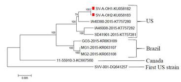 The dendrogram was constructed by using the neighbor-joining method in the MEGA software package, version 6.05 (www.megasoftware.net). Bootstrap resampling (1,000 replications) was performed, and bootstrap values are indicated for each node. Reference sequences obtained from GenBank are indicated by strain name and accession number. Scale bar represents 0.005 nucleotide substitutions per site.
The new rapid real-time RT-PCR is now available for detection of Senecavirus A RNA in clinical samples. Same-day testing can be completed if samples are received by 11 AM. Ideal sample types include vesicular scabs, swabs, or feces. The test costs $30.00.
To be PED, or not to be PED - that is the question!
Four 14-day-old nursing piglets were submitted for investigation of pre-weaning diarrhea. The farm had a history of porcine epidemic diarrhea virus (PEDV) infection on the premises. At necropsy, all piglets had similar changes. The stomachs were filled with casein curds. The small intestines were thin walled, flaccid and contained fluid yellow ingesta. The colon and rectum of each pig contained fluid yellow feces.
Small intestines of all four pigs exhibited mild to moderate segmental neutrophilic enteritis, with intralesional enteroadherent coccobacilli and intraluminal large bacterial rods. There was only very mild multifocal villous atrophy in intestinal sections of one pig.
Small intestinal tissues and content of each pig were subjected to real-time RT-PCR for PEDV, transmissible gastroenteritis virus (TGEV), and porcine deltacoronavirus (PDCoV). All four pig's samples were positive for PEDV, with Ct values ranging from 15 to 18, indicating a high viral load of PEDV. All samples were negative for TGEV and PDCoV. Next Generation Sequencing (NGS) indicated that the virus is the virulent strain of PEDV. Immunohistochemistry in this case performed at ISU VDL showed marked positive immunoreactivity of most enterocytes, compatible with a recent pathogenesis study that revealed: 1) positive IHC staining occurred at 12 hours post-inoculation, and 2) villous height / crypt depth ratios were normal at this early time point.
The lack of villous atrophy and attenuation of superficial villous enterocytes was an unusual finding in piglets with PED - All previous cases at the ADDL had severe villous atrophy and fusion of small intestinal villi. This case apparently represented a peracute case of PEDV infection, with evidence of marked widespread distribution of virus antigen prior to development of typical lesions of atrophic enteritis.
Meet the Staff: Virology Section
The ADDL is made up of five sections: Bacteriology, Central Receiving, Pathology, Serology, and Virology/Molecular Diagnostics/Avian Serology. Each section performs a vital role to the continuing success of the ADDL as your partner in veterinary diagnostics.
The Virology and Molecular Diagnostics Section has been led by Dr. Yan Zhang, DVM, PhD, DACVM since 2002. Eleven full-time staff members perform various virology and molecular assays, including virus isolation, fluorescent antibody tests, virus neutralization, ELISA, conventional and real-time PCR, and DNA sequencing. An average of 46,000 diagnostic virology tests and 30,000 molecular assays are performed each year.
A strong research and development team has improved the performance and testing capabilities of the Section. A number of new molecular assays and next generation sequencing has been introduced into the diagnostic testing repertoire.
Our highly trained staff are part of the National Animal Health Laboratory Network, partaking in several proficiency panels every year to ensure our competency to perform testing in the event of an outbreak of highly pathogenic avian influenza, foot-and-mouth disease, classical swine fever, or Newcastle disease.
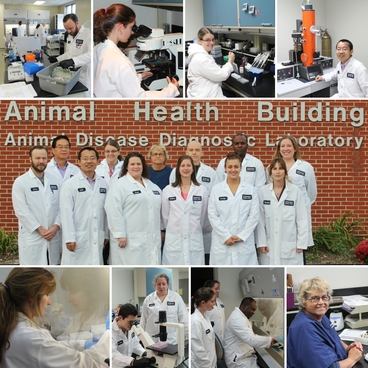 Center picture, Front row, L to R: Jason Herr, Leyi Wang, Kerri Lawrence, Brittany Garry, Jocelyn Black, Sandy Blackford. Back row, L to R: Yan Zhang, Jennifer Balough, Carolyn Gosnell, Troy Farrell, Olusegun Awe, Melanie Prarat
Bacteriology: Proficiency Test Update
The
Bacteriology Section successfully passed the NVSL Proficiency Tests for Johne’s
(Mycobacterium paratuberculosis) and
Contagious Equine Metritis (CEM) with scores of 100% for both NVSL proficiency tests.
The Johne’s proficiency test consisted of four tests, Pooled Fecal Culture,
Individual Fecal Culture, Pooled Fecal Direct PCR, and Individual Fecal Direct
PCR.
In addition to passing the CEM proficiency test, the Bacteriology
Section now has four approved analysts to conduct CEM testing. All analysts must successfully
complete a training course in the identification of Taylorella equigenitalius, the causative agent of CEM, at NVSL and
pass annually proficiency testing.
As a member of the Veterinary
Laboratory Investigation and Response Network (Vet-LIRN), the section
participated and scored a 100% in the “Salmonella in Dog Feces” proficiency
test organized by Vet-LIRN in collaboration with the Institute for Food Safety
and Health, Illinois Institute of Technology, and the Food and Drug
Administration.
Serology: Brucella canis Update
Currently, 3,473 dogs have been tested for Brucella canis at the ADDL. Of these animals, 29 have yielded
positive by the Indirect Fluorescent Antibody (IFA) test (0.8%). Among them, 21 samples (0.6%) have
been confirmed to be positive at 1:200 using Tube Agglutination Testing (TAT).
|