First Report: Seneca Valley Virus detected in Ohio
Ohio's first case of Seneca Valley Virus (SVV)
has been confirmed this month. A pig with vesicular lesions on its snout was identified and
samples were immediately collected and sent to ADDL and Plum Island Animal
Disease Center in Greenport, New York to rule out foreign animal diseases
(FADs), including foot-and-mouth disease (FMD). The samples were
FMD-negative but ADDL detected SVV nucleic acid present in the vesicular
and swab samples. Partial genome sequencing of the VP3 and VP1 structural
and 3D nonstructural proteins indicate 99% nucleotide identity with recent SVV strains
from the midwestern United States.
ADDL recently developed a RT-PCR that detects
two targets within the SVV genome. The
test is performed Monday-Friday with an anticipated turnaround time of 1-2
days. Fresh nasal or fecal swabs, serum
and feces are acceptable sample types.
The cost of the test is $30.00.
For more information, contact the ADDL at 614-728-6220 or animal@agri.ohio.gov.
Any swine having vesicular lesions on its snout,
around its coronary bands, or in both areas are suspects for FADs such as FMD
until determined otherwise by USDA APHIS Veterinary Services diagnostic
testing. During this SVV outbreak, it is important for producers and
veterinarians to continue reporting of pigs with vesicular lesions to ensure
rapid detection of potential foreign animal diseases, safeguard our
agriculture, and protect the health, quality, public confidence, and
marketability of our nation’s livestock and products.
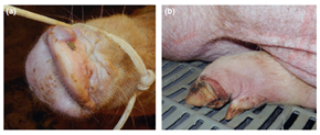 Vesicular disease associated with the presence of SVV. (a) Intact vesicle on the snout of an infected sow. (b) Erosive lesion bordering the coronary band in the left rear hoof of an affected sow. (Vannucci FA, Linhares DCL, Barcellos DESN et al. 2015. Identification and Complete Genome Sequence of Seneca Valley Virus in Vesicular Fluid and Sera of Pigs Affected with Idiopathic Vesicular Disease, Brazil. Transboundary and Emerging Diseases, 2015 Sept 7. PMID: 26347296.)
 Bovine abortions caused by Helcococcus ovis
A Holstein fetus aborted at 115 days in gestation along with
a placenta was submitted to ADDL. This was the fourth abortion in the herd over
an 8-month period. A moderate to heavy pure growth of Helcococcus ovis
was obtained from the placenta as well as from the lung and stomach contents of the fetus.
Microscopic examination of the allantochorion revealed severe necrosuppurative
placentitis with thrombosis, vasculitis and intralesional cocci. Lesions
in fetal tissues included moderate suppurative bronchopneumonia with
intralesional cocci, mild lymphohistiocytic myocarditis, mild lymphocytic
interstitial nephritis and also moderate neutrophilic rumenitis. Other tests
performed failed to detect additional pathogenic agents. Based on lesions in
multiple tissues, recovery of pure growth of H. ovis from two of
those tissues as well as from fetal stomach contents, and the exclusion of
other pathogens, a diagnosis of bacterial abortion associated with Helcococcus
ovis was made.
Helcococcus ovis is a Gram-positive, facultative
anaerobic coccus. It was originally isolated in 1999 from sheep. It is now
considered to be an emerging veterinary pathogen and has been reported as the
causative agent of bovine valvular endocarditis and metritis, pulmonary
abscesses in a horse, a goat and pleuritis and bronchopneumonia in sheep. H. ovis
was also isolated recently in the United Kingdom from the stomach contents of
an aborted bovine fetus, suggesting this agent as a potential causal pathogen
for the abortion. However, pathology from the aborted fetus or placenta was not
reported. To our knowledge, this is the first time that histological lesions
and isolation and identification of H. ovis were clearly linked to a bovine abortion.
|
 "Polio" (PEM) in a 200 pound dairy calf
A three
month old Holstein bull calf had a 2 day history of star gazing, opisthotonus,
lateral recumbency, muscle convulsions and blindness. The calf received two
injections of thiamine HCL the day prior to presentation and no clinical
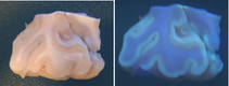
response was seen. Marked autofluorescence of the entire cortex of both
cerebral hemispheres was seen under UV light, consistent with
polioencephalomalacia (PEM). Microscopically, typical laminar neuronal
necrosis was observed in the cerebral cortical grey matter. Many cases of PEM can
usually be linked to feeding increased amounts of a high concentrate ration at
a rate that outpaces adaptation by rumen flora.
|
Virology
Porcine
reproductive and respiratory syndrome (PRRS) causes major problems for the
swine industry worldwide. At ADDL, we used whole genome sequencing to identify two novel strains of PRRS virus
from pigs experiencing severe clinical episodes of respiratory and reproductive
disease. Our
study highlights the importance of continued monitoring of PRRS virus using
whole genome sequencing. Reference: Wang L, Zhang Y. Novel porcine reproductive and respiratory syndrome virus strains in the United States with deletions in untranslated regions. Arch Virol. 2015 Sep 11. [Epub ahead of print] PubMed PMID: 26358265.
|