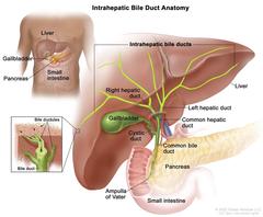
Anatomy of the intrahepatic bile duct; drawing shows the liver, intrahepatic bile ducts, right and left hepatic ducts, gallbladder, pancreas, and small intestine. An inset shows a cross section of a liver lobule with a network of bile ductules leading into a bile duct.
Anatomy of the intrahepatic bile ducts. Intrahepatic bile ducts are a network of small tubes that carry bile inside the liver. The smallest ducts, called ductules, come together to form the right hepatic bile duct and the left hepatic bile duct, which drain bile from the liver. Bile is stored in the gallbladder and is released when food is being digested.
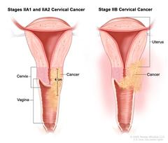
Stage II cervical cancer; drawing shows a cross-section of the uterus, cervix and vagina. In stages IIA1 and IIA2, cancer that is 4 cm is shown in the cervix and in the upper third of the vagina. In stage IIB, cancer is shown in the cervix, the upper two thirds of the vagina, and in the tissues around the uterus.
Stage II cervical cancer. Cancer has spread beyond the cervix but not to the pelvic wall or to the lower third of the vagina. In stages IIA1 and IIA2, cancer has spread beyond the cervix to the vagina. In stage IIA1, the tumor can be seen without a microscope and is 4 centimeters or smaller. In stage IIA2, the tumor can be seen without a microscope and is larger than 4 centimeters. In stage IIB, cancer has spread beyond the cervix to the tissues around the uterus.
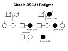
Pedigree showing some of the classic features of a family with a deleterious BRCA1 mutation across three generations, including transmission occurring through maternal and paternal lineages. The unaffected female proband is shown as having an affected mother (breast cancer diagnosed at age 42 y), female cousin (breast cancer diagnosed at age 38 y), maternal aunt (ovarian cancer diagnosed at age 53 y), and maternal grandmother (ovarian cancer diagnosed at age 49 y).
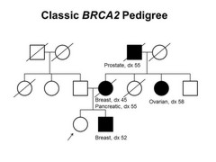
Pedigree showing some of the classic features of a family with a deleterious BRCA2 mutation across three generations, including transmission occuring through maternal and paternal lineages. The unaffected female proband is shown as having an affected brother (breast cancer diagnosed at age 52 y), mother (breast cancer diagnosed at age 45 y and pancreatic cancer diagnosed at age 55 y), maternal aunt (ovarian cancer diagnosed at age 58 y), and maternal grandfather (prostate cancer diagnosed at age 55 y).
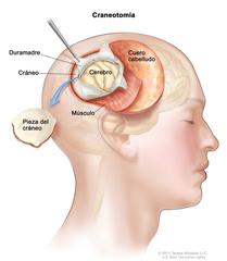
Dibujo de una craneotomía que muestra una sección del cuero cabelludo que se retiró hacia atrás para extraer un trozo del cráneo; se abrió la duramadre que cubre el cerebro para exponer el cerebro. También se muestra la capa de músculo bajo el cuero cabelludo.
Craneotomía. Se realiza una abertura en el cráneo y se extrae un trozo del cráneo para mostrar parte del cerebro.
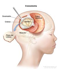
Dibujo de una craneotomía que muestra una sección del cuero cabelludo que se separó para extraer cráneo que se separó para extraer una pieza del cráneo; se abrió la duramadre que cubre el cerebro para exponer el cerebro. También se muestra la capa de músculo bajo el cuero cabelludo.
Craneotomía. Se realiza una abertura en el cráneo y se retira una pieza del cráneo para mostrar una parte del cerebro.
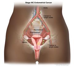
Stage IIIC endometrial cancer shown in a cross-section drawing of the uterus, cervix, fallopian tubes, ovaries, and vagina. Also shown are the lymph nodes in the pelvis and the aorta with nearby lymph nodes. Cancer is shown in the endometrium and myometrium of the uterus and in lymph nodes in the pelvis and near the aorta.
Stage IIIC endometrial cancer. Cancer has spread to lymph nodes in the pelvis and/or around the aorta (the largest artery in the body, which carries blood away from the heart).
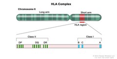
Human lymphocyte antigen (HLA) complex; drawing shows the long and short arms of human chromosome 6 with amplification of the HLA region, including the class I A, B, and C alleles, and the class II DP, DQ, and DR alleles.
HLA Complex. Human chromosome 6 with amplification of the human lymphocyte antigen (HLA) region. The locations of specific HLA loci for the class I B, C and A alleles and the class II DP, DQ, and DR alleles are shown.
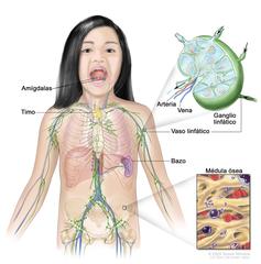
Sistema linfático; el dibujo muestra los vasos linfáticos y los órganos linfáticos, se incluye los ganglios linfáticos, las amígdalas, el timo, el bazo y la médula ósea. Un recuadro muestra la estructura interior de un ganglio linfático y de los vasos linfáticos adjuntos, con flechas que indican cómo circula la linfa (un líquido claro) hacia dentro y fuera del ganglio linfático. Otro recuadro muestra una vista ampliada de la médula ósea con células sanguíneas.
Anatomía del sistema linfático; se muestran los vasos linfáticos y los órganos linfáticos, se incluye los ganglios linfáticos, las amígdalas, el timo, el bazo y la médula ósea. La linfa (un líquido claro) y los linfocitos se desplazan a través de los vasos linfáticos hasta los ganglios linfáticos, donde los linfocitos destruyen las sustancias que son dañinas. La linfa entra en la sangre a través de una vena grande cerca del corazón.
Linfoma de Hodgkin en adultos en estadio III; el dibujo muestra un cáncer en grupos de ganglios linfáticos por encima y por debajo del diafragma, en el pulmón izquierdo y en el bazo. En un recuadro, se puede ver un ganglio linfático con un vaso linfático, una arteria y una vena. En el ganglio linfático se pueden ver las células de linfoma que contienen cáncer.
Linfoma de Hodgkin en adultos en estadio III. El cáncer se encuentra en uno o más grupos de ganglios linfáticos, por encima y por debajo del diafragma (a). En el estadio IIIE, el cáncer se encuentra en grupos de ganglios linfáticos por encima y por debajo del diafragma, y en un área u órgano cercanos fuera de los ganglios linfáticos (b). En el estadio IIIS, el cáncer se encuentra en grupos de ganglios linfáticos por encima y por debajo del diafragma (a), y en el bazo (c). En el estadio IIIS más el estadio E, el cáncer se encuentra en grupos de ganglios linfáticos por encima y por debajo del diafragma, en un área u órgano cercanos fuera de los ganglios linfáticos (b), y en el bazo (c).
Linfoma de Hodgkin en adultos en estadio I; el dibujo muestra un cáncer en un grupo de ganglios linfáticos por encima del diafragma. En un recuadro, se puede ver un ganglio linfático con un vaso linfático, una arteria y una vena. En el ganglio linfático se pueden ver las células de linfoma que contienen cáncer.
Linfoma de Hodgkin en adultos en estadio I. El cáncer se encuentra en uno o más ganglios linfáticos de un grupo de ganglios linfáticos. En el estadio IE (que no se muestra), el cáncer se encuentra en un área u órgano fuera de los ganglios linfáticos.
Linfoma de Hodgkin en adultos en estadio II; el dibujo muestra un cáncer en un grupo de ganglios linfáticos por encima y por debajo del diafragma. En un recuadro, se puede ver un ganglio linfático con un vaso linfático, una arteria y una vena. En el ganglio linfático se pueden ver las células de linfoma que contienen cáncer.
Linfoma de Hodgkin en adultos en estadio II. El cáncer se encuentra en dos o más grupos de ganglios linfáticos, y ambos están ya sea por encima (a) o por debajo (b) del diafragma.
Linfoma de Hodgkin en adultos en estadio IIE; el dibujo muestra un cáncer en un grupo de ganglios linfáticos por encima del diafragma y en el pulmón izquierdo. En un recuadro, se puede ver un ganglio linfático con un vaso linfático, una arteria y una vena. En el ganglio linfático se pueden ver células de linfoma que contienen cáncer.
Linfoma de Hodgkin en adultos en estadio IIE. El cáncer se encuentra en uno o más grupos de ganglios linfáticos por encima o por debajo del diafragma, y en un área u órgano cercanos fuera de los ganglios linfáticos.
Linfoma de Hodgkin en adultos en estadio IV; el dibujo muestra un cáncer en el hígado, el pulmón izquierdo y en un grupo de ganglios linfáticos por debajo del diafragma. También se pueden ver el cerebro y la pleura. En un recuadro, se puede ver en primer plano el cáncer que se disemina a través de los ganglios linfáticos y los vasos linfáticos hasta otras partes del cuerpo. En el interior de un ganglio linfático se pueden ver las células de linfoma que contienen cáncer. En otro recuadro se pueden ver células cancerosas en la médula ósea.
Linfoma de Hodgkin en adultos en estadio IV. El cáncer se encuentra fuera de los ganglios linfáticos a través de uno o más órganos (a); o fuera de los ganglios linfáticos en un órgano y se diseminó hasta ganglios linfáticos muy lejos de ese órgano (b); o está en el pulmón, el hígado o la médula ósea.
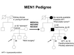
Pedigree showing some of the features of a family with a deleterious MEN1 mutation across four generations, including transmission occurring through paternal lineage. The unaffected male proband is shown as having an affected sister (self-report of neck surgery confirmed upon review of medical records to be hyperparathyroidism diagnosed at age 18 y, parathyroidectomy, and pituitary adenoma), father (self-report of stomach cancer confirmed upon review of medical records to be gastrinoma diagnosed at age 45 y), and paternal grandmother (suspected hyperparathyroidism and/or pancreatic tumor).
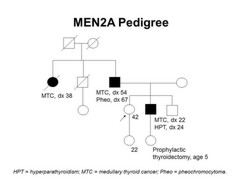
Pedigree showing some of the classic features of a family with a deleterious RET mutation across four generations, including transmission occurring through paternal lineage. The unaffected female proband is shown as having an affected brother (medullary thyroid cancer diagnosed at age 22 y and hyperparathyroidism diagnosed at age 24 y), father (medullary thyroid cancer diagnosed at age 54 y and pheochromocytoma diagnosed at age 67 y), and paternal aunt (medullary thyroid cancer diagnosed at age 38 y).
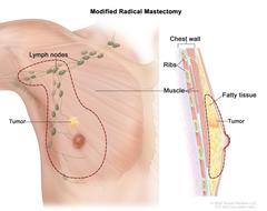
Modified radical mastectomy. The drawing on the left shows the removal of the breast, most or all of the lymph nodes under the arm, the lining over the chest muscles and sometimes part of the chest wall muscles. The drawing on the right shows a cross-section of the breast including the chest wall (ribs and muscle), fatty tissue, and the tumor.
Modified radical mastectomy. The dotted line shows where the entire breast and some lymph nodes are removed. Part of the chest wall muscle may also be removed.
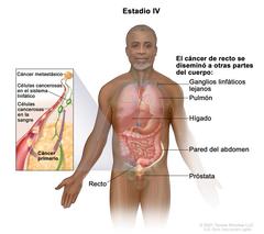
Cáncer de recto en estadio IV; el dibujo muestra otras partes del cuerpo hacia donde se puede diseminar el cáncer de recto, como los ganglios linfáticos, el pulmón, el hígado, la pared del abdomen y el ovario. En el recuadro se observa el cáncer que se está diseminando a través de la sangre y los ganglios linfáticos hacia otras partes del cuerpo.
Cáncer de recto en estadio IV. El cáncer se diseminó a través de la sangre y los ganglios linfáticos hacia otras partes del cuerpo, como el pulmón, el hígado, la pared del abdomen o el ovario.
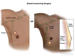
Cirugía para preservar la mama; el dibujo muestra la extirpación del tumor y los ganglios linfáticos de la axila.
Cirugía para preservar la mama. Las líneas de puntos muestran el área que contiene el tumor que se extirpa y algunos de los ganglios linfáticos que se pueden extirpar.