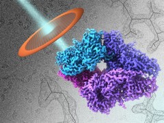
Artistic illustration of density map of the enzyme beta-galactosidase (beta-gal) determined by cryo-electron microscopy at 2.2 angstrom resolution. An essential enzyme in the human body, beta-gal is commonly used in molecular biology as a reporter marker to monitor gene expression. The foreground shows the overall shape of the molecule (right) and an electron beam striking a specimen grid (left), while the background shows images of single molecules (upper left) and the atomic contours of amino acids in the structure (lower right).
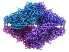
2.2 angstrom resolution electron density map of beta-galactosidase (beta-gal) as determined by high-resolution cryo-electron microscopy (cryo-EM). An essential enzyme in the human body, beta-gal is commonly used in molecular biology as a reporter marker to monitor gene expression. The map is colored by protein subunit. As of October 2015 it is the highest resolution structure determined by single-particle cryo-EM to date.
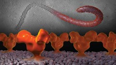
Cryo-electron tomographic image of a virus-like particle displaying Ebola virus surface spikes (top) and visualization of their three-dimensional structure (bottom).